Iron Deficiency Without Anemia: Fatigue, Ferritin, and Finding Balance
Iron deficiency can cause fatigue, brain fog, and restless legs even without anemia. Learn why ferritin often outperforms hemoglobin, how diet and traditional practices influence absorption, who may benefit from testing, and why too much iron can be harmful.
·10 min read
This content is for informational purposes only and does not constitute medical advice. Always consult a qualified healthcare provider before starting, stopping, or changing any supplement or medication regimen.
Iron Deficiency Without Anemia: Fatigue, Ferritin, and Finding Balance
Iron is best known for preventing anemia, yet research suggests many people experience iron-related symptoms well before hemoglobin drops. Fatigue, brain fog, hair shedding, brittle nails, and restless legs are frequently reported in non-anemic iron deficiency (NAID), a state in which iron stores are low but blood counts still appear “normal.” This article explores how to recognize NAID, why ferritin often outperforms hemoglobin as an early marker, how diet and tradition influence absorption, who may benefit from testing, and why more iron is not always better.
Non-Anemic Iron Deficiency: More Than Low Hemoglobin
NAID occurs when iron stores are depleted (often reflected by low ferritin) while hemoglobin remains within reference ranges. Symptoms may include fatigue, cognitive sluggishness, reduced exercise capacity, hair shedding, and restless legs. Menstruation, endurance training, frequent blood donation, pregnancy, gastrointestinal conditions that limit absorption (e.g., celiac disease, inflammatory bowel disease), heart failure, and chronic kidney disease are common contexts in which NAID may appear.
Treating low iron stores without anemia may reduce fatigue in menstruating adults. Randomized trials report modest improvements in fatigue when ferritin is low and anemia is absent (Evidence: moderate; RCTs and meta-analyses, e.g., BMJ 2003 and 2012).
Iron deficiency may impair cognition, attention, and memory even without anemia, especially in adolescents and people who menstruate (Evidence: moderate; systematic reviews suggest benefit of repletion on select cognitive tasks).
In heart failure, correcting iron deficiency (often intravenous in trials) may improve symptoms and quality of life regardless of anemia status (Evidence: strong; large RCTs such as FAIR-HF and CONFIRM-HF).
Ferritin: A Better Early Marker Than Hemoglobin Alone
Hemoglobin reflects red blood cell levels; it typically falls late in iron deficiency. Ferritin, a storage protein, declines earlier and generally tracks total body iron stores.
Ferritin is considered the best single laboratory indicator of iron stores in the absence of inflammation (Evidence: strong; WHO guidance and hematology reviews). However, ferritin rises with inflammation, liver disease, and metabolic conditions; in those cases, pairing ferritin with transferrin saturation (TSAT) and inflammatory markers (e.g., CRP) improves accuracy (Evidence: strong; clinical guidelines).
Relying on hemoglobin alone may miss symptomatic iron deficiency (Evidence: strong; diagnostic guideline consensus).
Heme vs. Non-Heme Iron: Why Your Plate Matters
Dietary iron comes as heme (primarily from animal foods) and non-heme (mostly from plants and fortified foods). Absorption differs substantially.
Heme iron is absorbed more efficiently and is less affected by other meal components (Evidence: strong; decades of absorption research).
Non-heme iron absorption varies widely and is enhanced by vitamin C–rich foods and depressed by phytates (in grains/legumes), polyphenols (tea/coffee), and calcium consumed with the same meal (Evidence: strong; controlled feeding studies and reviews).
Practical applications:
Pair legumes, whole grains, nuts/seeds, and leafy greens with vitamin C sources (citrus, bell pepper, strawberries, tomato) to support non-heme absorption (Evidence: strong; controlled trials of ascorbic acid and non-heme iron).
Soaking, sprouting, fermenting, and sourdough leavening may reduce phytate and modestly increase iron bioavailability in plant-based foods (Evidence: moderate; food science and human absorption studies).
Brewing tea or coffee away from iron-rich meals may help limit inhibition of non-heme iron uptake (Evidence: moderate; human absorption studies).
Traditional Foodways: Cast Iron, Organ Meats, and Ayurvedic Perspectives
Many culinary traditions feature iron-dense foods and techniques that naturally support iron status.
Cooking in cast-iron pans can increase the iron content of acidic, moist dishes; community trials using iron cookware have reported improvements in iron status in at-risk groups (Evidence: moderate; randomized and community studies in low-iron settings).
Traditional iron-rich foods include organ meats (liver), red meat, shellfish (e.g., clams, oysters), small fish eaten whole, and blood-based dishes; among plants, lentils, chickpeas, soy, blackstrap molasses, pumpkin seeds, sesame, amaranth, and dark leafy greens contribute non-heme iron (Evidence: strong for iron content; moderate for absorption in real-world diets).
Ayurveda has long used iron preparations such as Loha Bhasma (incinerated/processed iron) for conditions resembling weakness or pallor. Limited modern studies suggest hematinic effects, but concerns remain about standardization, bioavailability, and potential contamination with heavy metals if not prepared under strict quality controls (Evidence: traditional for use; emerging for modern efficacy/safety data). Individuals often look to food-first strategies and medically supervised products where quality is assured.
Restless Legs Syndrome (RLS) and Brain Fog: The Iron Link
Low ferritin is frequently observed in RLS, and iron therapy may reduce symptom severity in select cases (often when ferritin is low-to-borderline). Meta-analyses and guideline statements support iron repletion—particularly intravenous formulations in clinical settings—when iron stores are low (Evidence: moderate to strong; Cochrane review and sleep medicine guidelines).
Cognitive complaints and “brain fog” may relate to iron’s roles in myelination, neurotransmitter synthesis, and mitochondrial function. Trials and systematic reviews indicate that iron repletion can improve specific cognitive domains in iron-deficient individuals (Evidence: moderate).
Who May Benefit from Iron Testing
Population-wide screening is debated, but targeted assessment is common in groups with higher risk of iron depletion or symptoms suggestive of NAID. Research and guidelines suggest testing may be considered for:
Individuals with unexplained fatigue, reduced exercise capacity, hair shedding, brittle nails, pica, or restless legs, especially when risk factors are present (Evidence: moderate; RCTs linking repletion and symptom improvement, expert consensus).
People who menstruate—particularly with heavy or prolonged bleeding—or in the peripartum period (Evidence: strong; obstetric/gynecologic guidelines).
Endurance athletes and high-volume trainers, who may experience iron losses via foot-strike hemolysis, sweat, gastrointestinal microbleeds, and inadequate intake (Evidence: moderate; sports medicine reviews).
Those with gastrointestinal disorders affecting absorption (e.g., celiac disease, inflammatory bowel disease) or after bariatric procedures (Evidence: strong; gastroenterology guidelines).
Frequent blood donors (Evidence: strong; transfusion medicine guidance).
Individuals with chronic kidney disease or heart failure, where iron deficiency is prevalent and clinically relevant (Evidence: strong; nephrology and cardiology trials/guidelines).
When pursued, evaluation commonly includes ferritin, serum iron, total iron-binding capacity or transferrin, and TSAT, with awareness that inflammation can distort ferritin. Clinicians may also consider B12, folate, thyroid function, and markers of inflammation depending on history and exam.
Iron Overload: When “More” Becomes Risky
Iron is essential yet redox-active; in excess, it can catalyze oxidative injury. A small but significant portion of the population—particularly individuals of Northern European ancestry—carry HFE gene variants that predispose to hereditary hemochromatosis.
Hereditary hemochromatosis is characterized by elevated TSAT and ferritin, progressive tissue iron accumulation, and potential injury to the liver, heart, endocrine organs, and joints (Evidence: strong; genetic and clinical cohort studies; hepatology guidelines).
Secondary iron overload can occur with repeated blood transfusions, some anemias, chronic liver disease, and excessive iron intake over time (Evidence: strong; clinical reviews).
Before starting iron supplements, research-informed practice emphasizes confirming deficiency and ruling out overload or occult blood loss. Unnecessary iron may worsen conditions like hemochromatosis or complicate infections (Evidence: strong; guideline consensus). In malaria-endemic regions, iron supplementation without disease control increased infection risk in some trials; with appropriate malaria management, risk appears mitigated (Evidence: moderate; RCTs and systematic reviews).
Western and Eastern Views: Meeting in the Middle
Western biomedicine quantifies iron compartments (storage, transport, functional) and uses biomarkers like ferritin and TSAT to identify shortfalls or excess. Eastern traditions, including Ayurveda, emphasize digestive fire (agni), nourishment (rasa dhatu), and constitution (prakriti), with classical preparations (e.g., Loha Bhasma) aimed at restoring vitality. A practical bridge is to emphasize whole-food, well-prepared meals that enhance bioavailability (soaking/fermentation, vitamin C pairing), mindful use of traditional cookware, and individualized testing to clarify status before considering concentrated preparations.
Bottom Line
Iron deficiency can impair energy, cognition, and sleep quality even when hemoglobin is “normal.” Ferritin is typically a better early marker than hemoglobin alone, especially when interpreted alongside TSAT and inflammatory markers (Evidence: strong).
Dietary patterns and preparation matter: heme iron absorbs efficiently; non-heme iron benefits from vitamin C and traditional techniques that lower phytate, while tea/coffee and calcium taken with meals can inhibit absorption (Evidence: strong to moderate).
Cookware and cuisine count: cast-iron pans and iron-rich traditional foods may support iron status; Ayurvedic preparations reflect long-standing practice but require careful quality oversight in modern contexts (Evidence: moderate/traditional).
Not everyone benefits from more iron. Testing is often considered in higher-risk groups and in those with compatible symptoms, while screening for overload (e.g., hemochromatosis) protects against harm (Evidence: strong).
Personalized assessment helps determine whether low iron stores are contributing to symptoms—or whether another explanation better fits the picture.
References (selected)
Vaucher P, Druais P-L, Waldvogel S, Favrat B. Effect of iron supplementation on fatigue in nonanemic menstruating women with low ferritin: randomized trial. BMJ. 2012;345:e5843.
Verdon F, et al. Iron supplementation for unexplained fatigue in non-anaemic women: double blind randomized trial. BMJ. 2003;326:1124.
Anker SD, et al. Ferric carboxymaltose in patients with heart failure and iron deficiency (FAIR-HF). N Engl J Med. 2009;361:2436–2448. Ponikowski P, et al. CONFIRM-HF. Eur Heart J. 2015;36:657–668.
World Health Organization. Serum ferritin concentrations to assess iron status in individuals and populations. 2020.
Camaschella C. Iron-deficiency anemia. N Engl J Med. 2015;372:1832–1843.
Hurrell R, Egli I. Iron bioavailability and dietary reference values. Int J Vitam Nutr Res. 2010;80:257–265. Cook JD, Reddy MB. Am J Clin Nutr. 2001;73:93–98.
Adish AA, et al. Use of iron pots … improves iron status in Ethiopian children. Lancet. 1999;353:712–716.
Trotti LM, et al. Iron for the treatment of restless legs syndrome. Cochrane Database Syst Rev. 2019;CD007834. American Academy of Sleep Medicine guideline update, 2021.
Falkingham M, et al. The effects of iron supplementation on cognition in children and adults: systematic review and meta-analysis. Am J Clin Nutr. 2010;92:126–135.
Adams PC, Barton JC. Hemochromatosis. N Engl J Med. 2010;362:190–201.
Peeling P, et al. Iron considerations for the athlete: a narrative review. Sports Med. 2008;38:991–1006.
Sazawal S, et al. Effects of routine prophylactic supplementation with iron and folic acid on admission to hospital and mortality in preschool children in a high malaria transmission setting: cluster RCT. Lancet. 2006;367:133–143.
This content is for informational purposes only and does not constitute medical advice. Always consult a qualified healthcare provider before starting, stopping, or changing any supplement or medication regimen.
## Why You Can Feel Iron-Deficient Without “Anemia”
If you feel [persistently tired](/compare/chronic-fatigue-syndrome-me-cfs), unfocused, or plagued by restless legs but your blood count looks “norm
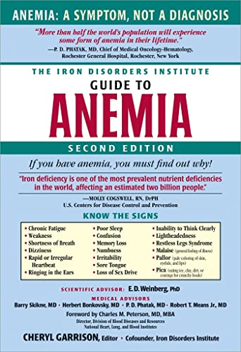
Iron Deficiency Anemia. . ## Overview
**Iron deficiency anemia (IDA)** is a common form of anemia that occurs when the body does not have enough iron to produce adequate amounts of hemoglobin, the oxygen-carrying component of red blood cells. As iron stores decline, red blood cell production becomes impaired, and tissues ma
Iron Panel. Iron Studies, Serum Iron Test. ## Overview
An **iron panel** is a group of laboratory tests used to evaluate how iron is being carried, stored, and used in the body. It is commonly ordered when clinicians are assessing possible **iron deficiency**, **iron overload**, unexplained fatigue, certain types of anemia, chronic inflammat
Ferritin Test. Iron Storage Test, Serum Ferritin Test. ## Overview
A **ferritin test** is a laboratory blood test that measures the concentration of **ferritin**, a protein that stores iron inside cells and releases it when the body needs it. Because ferritin reflects, to a significant extent, the body’s **iron reserves**, the test is widely used when
Essential Minerals for Optimal Health: Roles, Recommended Intake, Food Sources, Deficiency Signs, and Safe Supplementation. If you’re wondering which minerals actually matter, how much you need, and whether supplements are worth it, you’re not alone. Essential minerals for optimal health underpin everything from bone strength and energy production to thyroid balance and immune defense. This guide clarifies what each mine